
Why do Newborns Get Jaundice
Many infants, especially those born prematurely, develop jaundice. It happens when bilirubin—a substance released when your red blood cells break down—accumulates in the bloodstream. Infants are prone to jaundice, as their livers are not mature enough to remove the excess bilirubin from the blood.
The good news? Jaundice is often harmless and disappears on its own. However, it’s important to watch for the warning signs and see a pediatrician in Navi Mumbai if you notice unusual symptoms.
What Causes Jaundice in Babies?
Let’s understand the most common causes of jaundice in newborns.
1. Inadequate Feeding
Breastfed or formula-fed babies can both develop jaundice in the early weeks of birth due to inadequate feeding. If your baby doesn’t get enough milk, they may not defecate regularly, leading to bilirubin buildup in the blood.
You may need to talk to a lactation consultant or visit a pediatrician to discuss ways to improve feeding. Medical intervention, such as phototherapy, is recommended when bilirubin levels are 15-20 mg/dL or higher.
2. Breast Milk Jaundice
A chemical in the mother’s breastmilk can also cause jaundice in infants. This happens when the substances released in the breastmilk interfere with the bilirubin breakdown process. It usually starts a week after birth and may last up to 2-3 weeks or sometimes longer. Although breastmilk jaundice is harmless, it’s still best to keep in touch with a pediatrician and monitor bilirubin levels.
3. Babies have More Red Blood Cells
Babies have more red blood cells when they are in the uterus. These are like little oxygen tanks that compensate for the low oxygen levels in the womb. When they are born, they start breathing and no longer need such high levels of red blood cells.
As a result, these cells start breaking down rapidly, releasing a considerable amount of bilirubin. This, combined with irregular bowel movements, leads to excessive bilirubin in the baby’s blood, increasing their risk of jaundice.
4. Blood Group Incompatibility
When the baby’s and mother’s blood groups mismatch, the jaundice risk in the baby increases. When a baby is in the uterus, their blood can sometimes get mixed with that of its mother. If their blood groups are incompatible, the mother’s immune system might perceive it as a foreign object and create antibodies against it. These antibodies can cross the placenta and lead to the rapid destruction of the baby’s red blood cells, leading to more bilirubin production and a higher risk of jaundice.
Is Jaundice Serious?
Jaundice, in many babies, is harmless and often resolves within a few days without requiring any medical care. However, it’s always important to watch for extremely high bilirubin levels or uncommon symptoms. Here’s what may warrant prompt medical attention:
- Poor feeding
- Lethargy
- Excessive sleeping
- Yellowing in the legs and feet
Babies who develop jaundice immediately after birth may require admission to the NICU in Navi Mumbai, as it may indicate blood group incompatibility, infection, or rapid red blood cell destruction. Extremely high bilirubin can affect the baby’s brain, which is why doctors prefer regular monitoring to prevent complications.
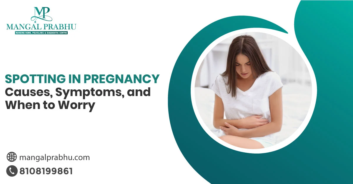
Spotting in Pregnancy: Causes, Symptoms, and When to Worry
Even a few drops of blood in your underwear can seem scary during pregnancy. Women often rush to the maternity hospital in Navi Mumbai to ensure their baby’s safety. The good news is that spotting, especially in the early months of pregnancy, doesn’t always mean danger.
That said, any form of bleeding during pregnancy must always be reported to a gynecologist, as it can indicate serious issues, such as a miscarriage, ectopic pregnancy, and other complications. In this post, we’ve discussed spotting in pregnancy, common causes, and when it requires medical attention.
Spotting and Bleeding Explained
Spotting means light bleeding, often just a few drops of blood that are visible, but not too much to soak through your underwear. Bleeding, however, requires pads or tampons. The color can be pink, red, or brown, and it usually lasts a couple of hours to a few days. Many women reporting spotting during pregnancy go on to have a safe pregnancy and an absolutely normal baby.
Common Causes of Spotting
The most common duration for spotting is the first 12 weeks of pregnancy. Here are the common causes:
Implantation Bleeding: In the early days, around 6-12 days after conception, the fertilized egg gets implanted into the uterus, causing implantation bleeding in some women. This is considered normal, given that the bleeding is light, lasts 1-2 days, and occurs with mild or no pain.
Hormonal Shifts: During pregnancy, hormonal changes in your body can make the cervix more sensitive. This makes light bleeding, especially after physical stress, internal examination, or sex, normal.
Cervical Issues: Problems with the cervix, such as cervical infection, polyps, or cervical erosion, can lead to bleeding during pregnancy. They require medical attention.
Placental Abruption: Detachment of the placenta from the uterus can cause heavy bleeding, which often occurs with heavy abdominal pain. Besides, placenta previa, a condition in which the placenta is attached deep down in the uterus, covering the cervix (partially or fully), can cause bleeding. Both issues are serious and require close observation.
Miscarriage or Ectopic Pregnancy: Although less common with first-trimester spotting, especially without other symptoms, miscarriage or ectopic pregnancies are a possible pregnancy complication. They occur with light bleeding that gradually becomes heavier and is accompanied by painful cramps.
When is Spotting Concerning During Pregnancy?
Although some bleeding during pregnancy doesn’t mean harm, it’s best to report it to a gynecologist in Navi Mumbai (no matter how brief or light it seems). This helps your gynecologist determine whether further tests are needed or if you require medication or monitoring.
What matters more is the timing of the bleeding. Spotting in early pregnancy can be implantation bleeding, but if the same occurs later in the second or third trimester, it can be concerning. Here are the signs you shouldn’t ignore:
- Heavy bleeding that requires pads
- Blood clots
- Dizziness
- Fever or chills
- Severe abdominal cramps
Besides, bleeding that looks like menstrual bleeding is not considered normal during pregnancy. See a gynecologist immediately if you experience these symptoms. Further tests, including blood tests or ultrasounds, may be needed to identify the cause.
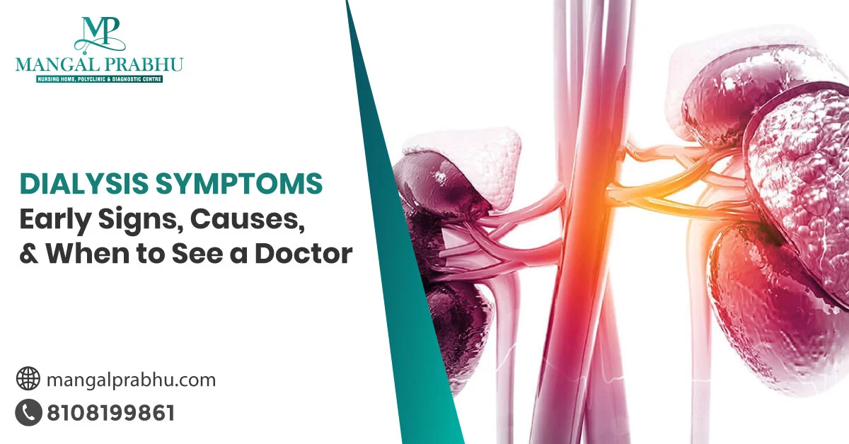
Dialysis Symptoms: Early Signs, Causes, and When to See a Doctor
When your kidneys do not filter the waste and excess fluid from your blood effectively, a nephrologist in Navi Mumbai might recommend dialysis. The symptoms that indicate a need for treatment can start much before your first dialysis session. They are often overlooked as they overlap with other non-alarming issues. The key is to keep an eye on your symptoms and report anything that seems unusual to your doctor.
Overview of Dialysis
The main job of a healthy kidney is to remove waste, fluids, and toxins from your blood, keeping it clean. Your kidneys also balance electrolytes and hormones. Chronic kidney diseases or kidney failure can result in a drop in its function, making treatment necessary.
That’s where dialysis comes into the picture. It’s a common procedure that mimics the function of your kidneys. The number of sessions required for dialysis weekly depends on whether it’s hemodialysis or peritoneal dialysis. Note that dialysis isn’t a cure for kidney disease. It’s rather a procedure to stabilize your body’s vital functions until you find a new kidney.
Signs You Need Dialysis
Initially, symptoms of a potential kidney disease may be too mild to seem concerning. Many people mistake the subtle signs for everyday stress or fatigue. However, as the disease progresses, the symptoms may become more pronounced. Here are the common signs you shouldn’t ignore.
Urination Issues
You notice changes in the urination pattern when protein starts leaking into the urine. Common signs include little urine output or complete cessation. Foamy urine or dark-colored urine. Or, a need to urinate frequently at night, also called nocturia.
Constant Fatigue and Weakness
When kidneys no longer filter toxins, these waste substances may build up in your blood, making your brain and other organs sluggish. Additionally, the kidneys also play a part in making red blood cells. So, when their function drops, you might have a higher risk of anemia, which can make you weak and feel low on energy despite rest.
Swelling in the Feet, Ankles, and Hands
When excess fluid isn’t removed from your body, it pools in your legs, hands, ankles, and other body parts, making them swell. If you notice a puffy face, swollen legs, or unusual tightness in the finger rings, these are all signs your kidneys might be struggling.
Nausea or Vomiting
Accumulation of waste substances in your blood can affect your taste and digestive function, which might result in loss of appetite, metallic taste, or nausea or vomiting.
Other Symptoms
Many patients who are referred to the dialysis center in Navi Mumbai experience one or more of these symptoms:
- Shortness of breath
- Constant itching
- Muscle cramps
- Difficulty concentrating
- Dizziness
- Pale skin
- Chest pain or discomfort
See a healthcare provider immediately if you experience chest pain, excessive swelling, or low urine output or no urine in the last 12-24 hours.
Dialysis is not scary. If a nephrologist recommends dialysis, it’s only because the benefits outweigh the risks. Timely diagnosis and treatment can prevent life-threatening complications associated with kidney diseases.

What Is Giardiasis? Causes, Symptoms & Treatment
Giardiasis is a parasitic infection that affects your small intestine, causing stomach cramps and diarrhea. This tiny, microscopic parasite called Giardia is found in every part of the world, and especially thrives in places with poor sanitation. It enters your body through contaminated food or water and finds its place in your small intestine, where it multiplies and affects your nutrient absorption.
With proper treatment of giardiasis in Navi Mumbai, it’s possible to recover fully from this parasitic infection. What’s more important is staying aware of the causes, symptoms, and treatment options so you can take preventive steps and quick action if you or someone close to you gets infected.
Causes
Giardia lives in the small intestines and eventually passes through poop. Before it exits your body, it turns into a protective shell, which is hard enough to survive in lakes, pools, spas, grounds, soil, and other unclean surfaces.
When someone swallows these cysts through water or food, these shells get into their intestines and break open. The germs living inside come out. The cycle continues. Here’s what increases the risk of infection:
- Eating food washed with contaminated water
- Drinking contaminated water
- Living in areas with poor sanitation
- Not washing hands after using the toilet
- Person-to-person contact (changing children’s diapers)
- Traveling to places with poor hygiene practices
Symptoms
Not everyone who gets infected with Giardiasis develops obvious symptoms. However, the parasite living in their small intestines can still infect others once it’s released. For people who do experience symptoms, here’s what happens:
- Loose watery stools
- Foul-smelling stools
- Bloating
- Gas
- Stomach cramps or abdominal pain
- Fatigue
- Weakness
- Nausea or vomiting
Children are more vulnerable to infection, especially those who wear diapers or are in the potty-training phase. Those attending day care are also at an increased risk. Symptoms start 1-3 weeks after infection.
The cyst opens soon after being swallowed and releases germs inside your intestine, but the symptoms might appear later. The parasitic infection might take between 2 and 6 weeks to clear completely, sometimes longer.
Diagnosis and Treatment
Giardiasis is diagnosed with a stool test. A general physician in Navi Mumbai might order it if you have the above-listed symptoms. Sometimes, more than one stool test may be conducted, as the parasite doesn’t always appear in every stool sample.
Fortunately, the treatment for giardiasis is often quite effective. Once the test results confirm the infection, the doctor might recommend anti-parasitic medication to clear it. Make sure you complete the full course even if the symptoms disappear.
Giardiasis in children requires prompt treatment, as frequent watery stools can lead to dehydration. It’s important to follow a light diet plan for your children during the recovery. Make sure they are hydrated. Doctors may prescribe oral rehydration solutions to prevent dehydration.
Conclusion
Giardiasis might sound alarming, but it is usually a manageable infection, especially when diagnosed and treated properly. Simple steps, such as following proper hygiene, eating well-cooked and properly washed foods, and drinking safe water, can help prevent the infection.

Vaginal Dryness After Period: Causes, Symptoms & Treatment
Have you ever felt an itching sensation with mild irritation down there? Although it’s more common in postmenopausal women, younger women can get it too, especially right after their periods. To help you understand it better, we’ve compiled a detailed post that lists the causes, symptoms, and treatment for vaginal dryness in Navi Mumbai. Here’s a look.
What is Vaginal Dryness?
Vaginal dryness is exactly what it sounds like — dry vaginal walls due to a lack of enough lubrication. Natural lubrication in your vagina keeps the tissues down there moist. The lack of moisture in your vagina can lead to itchiness, tightness, and irritation, which can affect your everyday life, relationship, and even sleep.
What Causes Vaginal Dryness After Period?
Let’s explore the common causes of vaginal dryness.
Low Estrogen: Your estrogen levels drop temporarily before your period. Since estrogen plays an important part in keeping your vaginal walls moist and elastic, a dip in its level can cause vaginal dryness.
Using Pads: Wearing pads for long hours can cause friction, especially in hot weather. You might feel itchiness or dryness down there. Likewise, tampons can sometimes absorb vaginal moisture along with the menstrual blood, making your vagina dry after periods.
Harsh Soaps: Too much use of scented soap and cleansing products can affect the natural pH balance of your vagina, causing irritation and a burning sensation.
Dehydration: If your body doesn’t get enough fluids, it won’t produce natural lubrication, making vaginal dryness more likely before or after periods.
Sometimes, underlying health issues, such as hormonal imbalance and skin disorders, can lead to vaginal dryness. If it’s frequent and is accompanied by pain and itching, it’s worth getting checked by a gynaecologist in Navi Mumbai.
Symptoms of Vaginal Dryness
If you have vaginal dryness, you will likely experience:
- A feeling of itchiness, tenderness, or irritation in or around your vaginal area
- Discomfort in your vulva after using the bathroom and while sitting or walking
- Spotting or bleeding after sexual intercourse
- Pain and discomfort during sex
- Discomfort when wearing pads or tampons
Note that these symptoms are temporary and often resolve on their own once your estrogen levels return to normal.
Treatment for Vaginal Dryness
The good news is vaginal dryness after periods is manageable. The main line of treatment depends on the causes. Here’s what may help.
Estrogen Creams: You can use estrogen cream to increase estrogen levels in your intimate area and get relief from constant itchiness and dryness around your vagina. Before using it, consult a gynaecologist to learn its benefits and potential side effects.
Water-based Lubricant: A gentle water-based lubricant is considered safe for daily use, especially during intercourse, to prevent discomfort due to vaginal dryness.
Moisturiser: You can also use a vaginal moisturiser every 3-4 days to maintain moisture in your intimate area.
Stay Hydrated: If vaginal dryness occurs due to dehydration, drinking enough fluids will restore moisture. Aim for 8-10 glasses a day, especially during and after periods (unless your doctor advises otherwise).
Most women experience vaginal dryness for 2-4 days after their period ends. In most cases, vaginal dryness after periods is not considered harmful. It’s linked to hormonal fluctuations.

Is a Brain Tumour Curable or Not
Hearing the word “brain tumour” can be overwhelming. The first question people ask is “Is a brain tumour curable?” It depends largely on the tumour type, i.e. whether it’s malignant or benign. Malignant tumour means it’s cancerous, and is more likely to grow faster and spread into the surrounding areas. Benign tumours are easier to treat, as they grow slowly and stay in one place. Here’s what an oncologist in Navi Mumbai says about brain tumour types and their curability.
Types of Brain Tumour
Let’s take a look at the common types of cancerous and non-cancerous brain tumours.
- Chordomas: These are found on the base of your skull or at the end of your spine.
- Craniopharyngiomas: The tumours grow near the pituitary gland and are often difficult to treat because of their location near the vital structures of your brain.
- Astrocytoma: These are gliomas, the tumours that develop in glial cells. Astrocytomas can be benign or malignant, depending on the stage. They are mostly found in the cerebrum.
- Glioblastoma: These are a type of astrocytoma, but tend to grow aggressively.
- Gangliocytomas: These are also non-cancerous brain tumours that develop in your nerve cells, but are often rare.
- Meningiomas: They grow in the meninges, protective layers around your brain. They are mostly benign, but can be cancerous in rare cases.
Are Brain Tumours Serious?
Brain tumours, whether benign or malignant, can be serious, especially if they are located in areas that control vital functions. Your skull is a hard space that doesn’t allow for a tumour to grow.
So, when a tumour forms inside your brain, it will squeeze the structures in your brain. The symptoms of a brain tumour vary depending on the type and location of the growth, but most people experience these symptoms:
- Weakness
- Unable to walk properly
- Vision loss
- Speech difficulties
- Memory problems
Some people with brain tumours may never know they have one, as they don’t experience any symptoms. The tumours don’t grow or squeeze the brain either.
Can Brain Tumours be Cured?
The success rate of cancer treatment in Navi Mumbai depends on many factors. The most crucial of all is the type of tumours. Benign brain tumours are easier to treat, as they may not have spread to other parts of the brain. The location also affects the treatment.
If the tumour is located in sensitive parts that are responsible for controlling speech, vision, memory, and more, the surgery to remove it completely might be risky. Likewise, size plays a role here. Larger tumours might be more difficult to remove compared to the smaller, benign ones, which are less likely to recur after complete removal.
Other factors are patients’ age and overall health. Younger, healthier patients tend to respond well to the treatment compared to older adults with weakened immunity.
Conclusion
Some tumours are curable, others can’t be cured fully, but are treatable. Your healthcare provider will work with you to find a personalized and most appropriate treatment plan that helps you live a long, meaningful life.
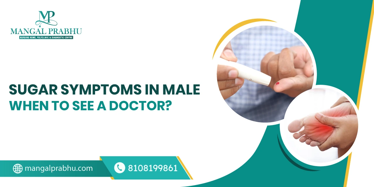
Sugar Symptoms in Male: When to See a Doctor
A spike in the blood sugar levels may not always produce the same symptoms in all individuals. Men, specifically, experience subtle signs, which become more pronounced later. In this post, we’ll take a look at the early warning signs of diabetes in men and when you need to schedule a visit to the multispeciality hospital in Navi Mumbai.
Understanding High Blood Sugar Levels
High blood sugar levels, called diabetes in medical terms, occur when your body doesn’t produce sufficient insulin or can’t use it efficiently. Insulin processes glucose by transferring it into your cells, where it can be stored or used for energy. When this hormone doesn’t function normally, glucose is stored in the blood, causing many health issues.
1. Erectile Dysfunction
Diabetic men are three times more likely to experience erectile dysfunction compared to non-diabetics. That’s because high blood sugar levels can damage their nerves and blood vessels, making it hard for men to get and keep an erection.
2. Frequent Urination and Excessive Thirst
When an excessive amount of glucose builds up in your blood, your kidneys might work harder to flush the sugar out of your system through urine. This leads to frequent urination. Since you are losing too much fluid through urine, you might feel thirsty frequently. Even if you have just had water, your mouth might feel dry.
3. You Feel Hungry Often
An obvious sign of diabetes in men is the constant feeling of hunger. When your body doesn’t get enough glucose, it will break down muscles and fat for energy. As a result, you will often feel hungry, and no matter how much you eat, you will lose weight. Unexplained weight loss or the desire to eat frequently can be the early signs of diabetes.
4. You have Blurry Vision
High blood sugar levels also affect your vision. It can cause swelling in the eye lens, as a result of which, your vision might be unclear or blurred. Getting your blood sugar levels under control will most likely restore your normal vision.
5. Wounds that Heal Slowly
High blood sugar levels can disrupt blood circulation, especially to the lower legs and feet. This, combined with nerve damage, can make it harder for the wounds to heal. Diabetic patients are often advised to see a general physician in Navi Mumbai if they develop sores or foot ulcers.
When left untreated for long, these can become infectious (especially in people with uncontrolled sugar). Although less likely, some cases require leg amputation to prevent the risk of the infection spreading.
6. Constant Fatigue
Glucose is supposed to travel to your cells so they can be used for energy. When they remain in your blood for too long, your body may not get enough energy, which can lead to constant fatigue. You might feel tired even after getting adequate rest. Another reason is frequent urination. It can, in some cases, lead to dehydration, which can leave you feeling exhausted.
These were the common signs of diabetes in men. If you believe your sugar levels in the blood are high, it’s important to see a general physician to prevent complications.

Diet for Appendicitis Patients: What to Eat and Avoid for a Smooth Recovery
Appendicitis is the inflammation of the appendix, a small organ attached to the large intestine. A surgery to remove the appendix might be necessary if the infection is severe.
According to general surgeons in Navi Mumbai, a crucial part of your recovery journey after an appendectomy is your diet. In this post, you’ll learn about the foods to eat and avoid during recovery. Here’s a look:
Foods to Eat after an Appendectomy
Your digestive system will be a little sensitive after surgery, so your diet should mainly include soft, easy-to-digest foods. Here’s a look at the recommended diet for appendicitis patients.
1. Clear Liquids
In the first few hours after surgery, your healthcare provider recommends a clear liquid diet. These are easy on your gut, keep you hydrated, and help your body transition smoothly. You can have fruit juices (without the pulp), broths, soups, herbal teas, coconut water, and more.
2. Soft Foods
When your doctor says it’s safe to start eating, you can introduce soft, bland, easily-digestible foods to your diet. Include foods that provide you with the essential nutrients while being gentle on your digestive tract. Examples include Khichdi, porridge, banana, mashed potatoes, stewed apples, and soft-boiled eggs.
3. Fiber-Rich Foods
Constipation is a common issue in people who have undergone an appendectomy. Pain meds with reduced physical activity can make constipation worse. To help ease and regulate your bowel movements, include fiber-rich foods in your diet. Examples include oatmeal, papaya, cooked carrots, spinach & other green, leafy vegetables, as well as whole wheat bread. That said, you shouldn’t start fiber soon after surgery. Introduce it gradually.
4. Protein-Rich Foods
Foods rich in protein strengthen your immunity and help with tissue repair. For smooth and quick healing, consider adding protein-rich foods like scrambled or boiled eggs, moong dal, fish, yogurt, and tofu (to name a few) to your diet.
What to Avoid After an Appendectomy
Not all foods are considered safe after appendix treatment in Navi Mumbai. It’s generally advised to avoid a diet that consists of hard-to-digest foods or foods packed with little to no nutrients. Here’s a list.
1. Spicy and Greasy Foods
Deep-fried and spicy foods are a big no-no after an appendectomy. These can irritate your digestive tract, cause acidity, and lead to indigestion. Examples include pakoda, samosa, spiced curry, and street foods.
2. Processed Foods
Chips, instant noodles, and creamy desserts are high in calories but lack nutritional value. Besides, they are hard on your digestive system.
3. Red Meat
Although red meat has a good amount of protein, it’s not easily digested and might also cause constipation and bloating, which can make your recovery uncomfortable.
4. Alcohol and Caffeine
Alcohol slows recovery and can interfere with your medication. It’s best to avoid it after an appendectomy. Likewise, caffeinated beverages can cause dehydration.
Conclusion
Appendectomy (the removal of an appendix) is the most effective treatment for appendicitis. Although having your appendix removed isn’t associated with long-term health risks, the surgery can cause discomfort, tenderness in the surgical area, and pain until you have healed. The right food won’t just speed up recovery, but make it comfortable.
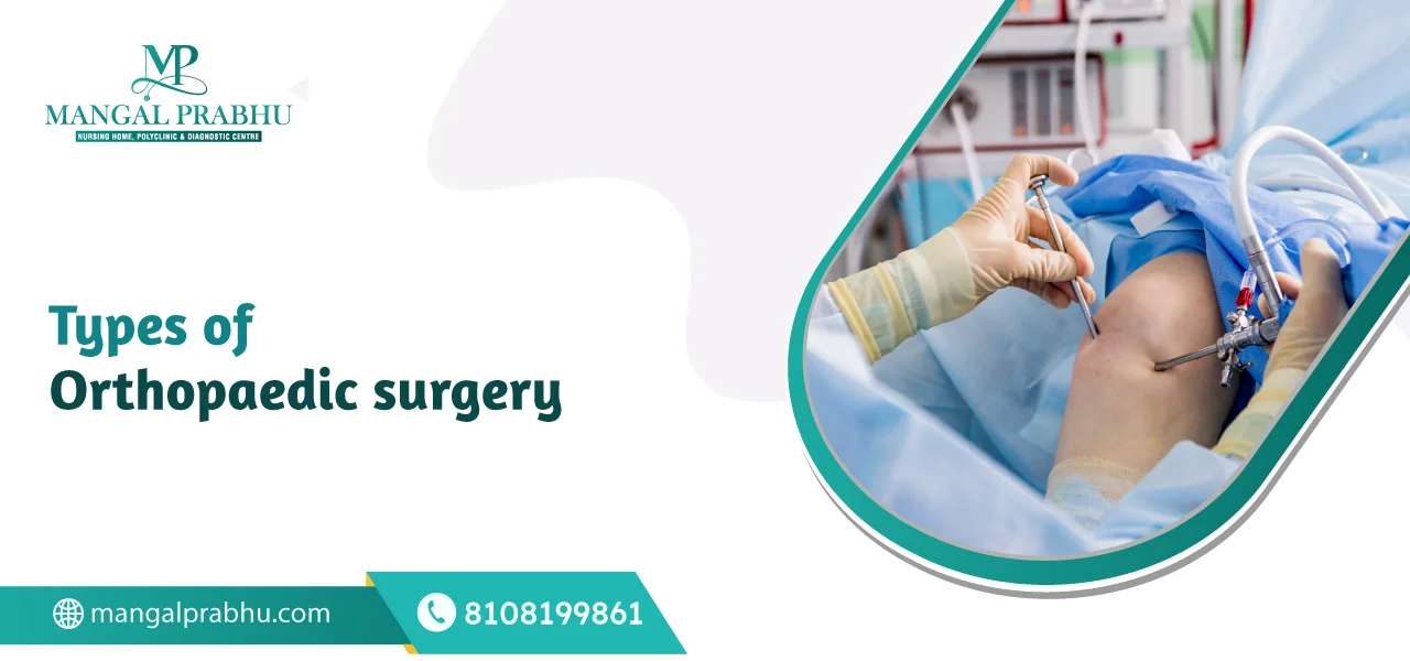
Types of Orthopedic Surgery
Treatments for conditions affecting your musculoskeletal system fall into the orthopedic surgery category. It involves your muscles, tendons, ligaments, joints, bones, nerves, and other interconnected structures. This branch of surgery helps people achieve stability, improved mobility, and a pain-free life. If you ever wonder about the types of procedures carried out in an orthopedic hospital in Navi Mumbai, this post is for you.
Common Types of Orthopedic Surgery
1. ACL Reconstruction
Do you hear a “pop” sound after sustaining a knee injury? Does your knee feel unstable? You might have suffered an Anterior Cruciate Ligament (ACL) injury. A torn ACL can make jumping, running, and even walking difficult. It requires surgical reconstruction, in which the ACL is replaced with a tissue graft.
2. Hip Replacement Surgery
Hip replacement is another orthopedic surgery that involves replacing the hip joint (ball or socket, or both). It’s recommended for patients with reduced mobility, debilitating hip pain, and difficulty performing routine tasks.
If you have rheumatoid arthritis, osteoarthritis, a hip fracture, or other medical issues with your hip joint that affect your joint stability, a hip replacement surgery can be an effective decision.
3. Knee Replacement Surgery
For severe arthritis that has limited your movements, either partially or entirely, knee replacement surgery can offer significant relief. With age, the cartilage that cushions your knee joints wears out, causing intense pain as your bones rub against each other with movement.
Knee replacement removes the damaged knee joint and replaces it with an artificial implant that improves mobility, reduces pain, and makes your knees feel more stable.
4. Knee Arthroscopy
An orthopedic surgeon in Navi Mumbai might recommend this minimally invasive surgical procedure for patients with a torn ACL, meniscal tears, and other minor knee joint issues.
In a knee arthroscopy, an arthroscope (a narrow device with a camera on one end) is inserted into your knee joint through a buttonhole-sized incision. The detailed picture of the inside of the knee is produced on a high-definition video monitor. It’s used for both diagnosis and treatment of joint issues.
5. Spinal Surgery
A spinal surgery is not usually recommended as the first line of treatment. However, if the consecutive treatments, such as rest, medication, and physiotherapy, do not help, surgery may be necessary. For spinal deformity, injuries or fractures that damage the spine, severe cases of slipped disc, infections, and spinal tumors, spinal surgery might be needed.
6. Shoulder Arthroscopy and Replacement
If your shoulder joints are not moving or functioning properly due to avascular necrosis, a shoulder fracture, rotator cuff tear, arthropathy, or osteoarthritis, a shoulder replacement surgery can be helpful. Your surgeon might first use shoulder arthroscopy to diagnose the extent of the damage. If necessary, they may replace the worn-out or dysfunctional shoulder joint with prosthetic implants.
7. Fracture Repair
For simple fractures, immobilizing the affected joint with splints or casts may be enough. For complex fractures, in which the broken bone pierces through the skin or is broken into several fragments, a more complex surgical approach may be used. The surgeon may use plates, screws, rods, pins, and other external fixators to keep the bone in place and offer stability as it heals.
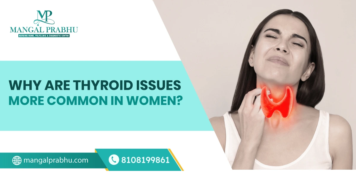
Why Are Thyroid Issues More Common in Women?
Certain medical illnesses are more prevalent in women than in men. Thyroid imbalance is one such example. According to the National Library of Medicine, women are 5-20 times more likely to develop thyroid disorders compared to men.
Thyroid issues affect your metabolism and might also interfere with pregnancy. The question is, why are most thyroid patients who visit a multispecialty hospital in Navi Mumbai women? Let’s find out.
What is Thyroid?
The thyroid is a small butterfly-shaped organ that regulates your metabolism, body temperature, mood, and growth. If it doesn’t function properly, it may produce either excessive or very little thyroid hormones. This can affect your hair health, mood, temperature, and periods.
Reasons Why Thyroid Issues are More Common in Women
1. Hormonal Fluctuations
A woman goes through hormonal fluctuations during puberty, menstruation, pregnancy, postpartum, and menopause. Estrogen is considered a vital hormone in regulating thyroid balance. Its imbalance can increase your risk of having thyroid dysfunction.
Besides that, the workload on your thyroid surges when you are pregnant. The increased demand can sometimes lead to thyroid disorders during pregnancy.
2. Autoimmune Response
Some thyroid problems, such as Graves’ disease, can affect your thyroid hormone. It can trigger an overproduction of the hormones, resulting in hyperthyroidism. Graves’ disease is an autoimmune disorder.
Many issues that affect thyroid production are linked to autoimmune disorders, a condition in which the immune system targets the body’s own healthy cells and tissues. Women tend to have a stronger immune system, which also makes them more prone to autoimmune disorders that can disrupt their thyroid function.
3. Genetic Factor
If you have a family history of thyroid issues, you are at an increased risk of getting them, as genetics affect your likelihood of developing certain illnesses. This risk is more prominent in women, as genes linked to autoimmunity are more active in women, making their bodies more susceptible to thyroid disorders.
4. Stress
Stress produces cortisol and adrenaline, the two hormones that help you deal with emergencies. However, when the stress is persistent or turns into a chronic health condition, it starts to interfere with your hormone function. It can also affect your thyroid gland.
Since women have a comparatively more active autoimmune system, consistent stress can worsen. Besides, excessive stress can cause inflammation, which can harm your thyroid gland.
5. Nutritional Deficiencies
For your thyroid gland to function normally, it requires certain nutrients, including Iodine, Selenium, Zinc, Vitamin D & B12, and Iron. If your diet doesn’t consist of foods rich in these nutrients, your risk of developing thyroid disorders increases.
Women are more likely to have nutritional deficiencies. For example, iron loss can occur during menstruation. During pregnancy, a woman’s nutritional requirement increases to support the growing baby. They are more susceptible to iron and iodine deficiencies around this time.
When their diet lacks the essential nutrients for thyroid production, they might develop hypothyroidism.Contact a thyroid specialist in Navi Mumbai to learn more about hyperthyroidism and hypothyroidism and how they affect your health.